![Collective Minds_logo.png]](https://help.collectiveminds.health/hs-fs/hubfs/Collective%20Minds_logo.png?height=22&name=Collective%20Minds_logo.png)
1. Windowing > Auto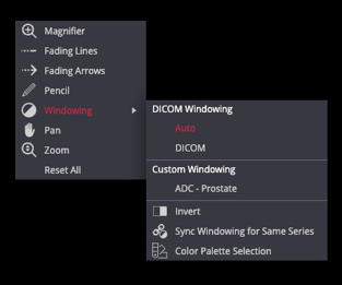
Shortcut: 0
Helps to automatically adjust the series window level and width. This tool is useful to correct a suboptimal MRI window and ADC maps
2. Preload Study or Series
To enhance scroll speeds and auto-synchronisation 
-
-
Click the cloud icon beside the study/series in the left panel.
-
Tip: Drag a series description into the viewport to auto-preload.
-
Preloading stores data temporarily in memory for smoother scrolling
-
3. Crosshair Tool
Enables synchronized navigation between series by selecting a point in one image that acts as a locator while referencing other planes. Useful for lesion localization in various planes. This tool may appear via the more options menu on smaller screens.
4. Measurement Tools
▫️ Line D
Measure distances, such as lesion size or organ dimensions, by drawing a straight line.
▫️Ellipse: Shift + E 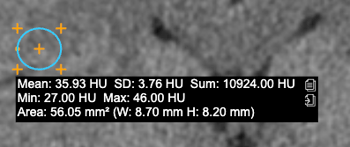
Draw an elliptical region of interest (ROI) to measure signal intensity values like HU, mean, standard deviation, Max, Min, and area.
Tip: Press E to erase lines or ROI.
▫️ TIC Tool (Time-Intensity Curve)
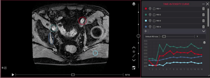
Useful in Dynamic Contrast-Enhanced (DCE) MRI to assess enhancement behaviour over time. Place regions of interest to generate a graph showing signal intensity changes during contrast phases.
A full preload of the dynamic series is required for this tool to function. Some Dynamic MRI series may be provided as separate series instead of a single large volume stack.
For studies with multiple DCE phases split across separate series:
-
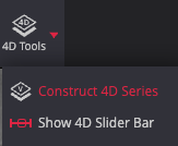
Try the 4D Tools > Construct 4D "virtual" series function to mark and combine series with matching descriptions into a larger virtual series stack. The virtual series requires a full study preload and, if successful, will appear as the last series in the study.
For more details, see here
5. Diffusion Weighted Imaging (DWI)
The b-value may be displayed in the top-left corner of the viewport, provided it is included in the DICOM metadata. This information varies between different MRI vendors, and values may also be included in the series description, for example.
Once preloaded, a DWI series containing multiple b-values may appear in the thumbnail panel as separated Phases, with each thumbnail representing a different b-value. Drag and drop a Phase thumbnail into the viewport to display the corresponding value series.
The 4D slider bar (shortcut: S) can also be used to navigate a preloaded DWI series stack. For DWI series from some vendors, creating a 4D series from the stack may further improve navigation and make it easier to review images across different b-values using the slider bar.
Windowing > Auto (Shortcut: 0)
Automatically adjusts the window level and width. This tool is useful for correcting suboptimal MRI windowing and improving the visibility of the DWI and ADC maps, for example.
6. DICOM Metadata Considerations
Advanced tools such as auto-linking, crosshair synchronization, diffusion b-values, and series merging depend on intact DICOM metadata. If images are in JPEG format, or if metadata has been altered or removed before upload, such viewer tools may not function correctly.